AI Model E(3)-Pose Enhances Prenatal Diagnosis by Tracking Fetal Movement
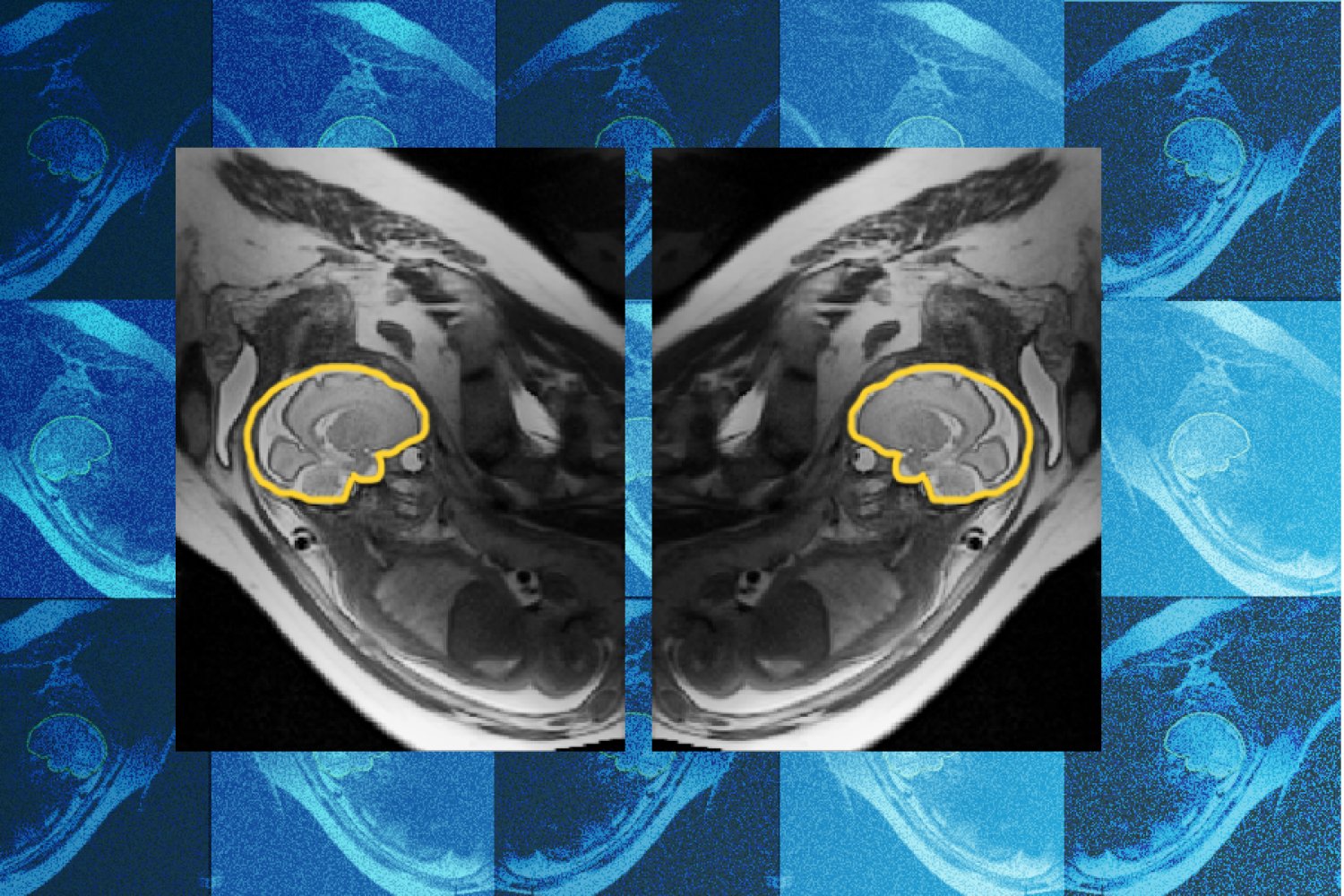
Researchers from MIT, Boston Children's Hospital, and Inria have developed a novel deep learning model named E(3)-Pose that significantly enhances the reliability of prenatal diagnoses by accurately imaging fetal heads even during movement. This innovation addresses a critical challenge in fetal Magnetic Resonance Imaging (MRI), where fetal motion can lead to incomplete or distorted scans, potentially causing missed or incorrect diagnoses of developmental abnormalities.
How E(3)-Pose Works
The E(3)-Pose model tackles the issue of fetal movement by integrating fundamental physics principles directly into its neural network architecture. Unlike models that learn physics from data, E(3)-Pose embeds concepts like the left-right symmetry of the fetal head. This approach allows the model to more effectively track the fetus's orientation and motion, ensuring that the 2D image "slices" captured during an MRI are consistently aligned. This consistency is crucial for reconstructing a high-resolution, volumetric 3D image of the fetal brain, which is essential for identifying subtle developmental anomalies.
Addressing a Clinical Pain Point
The development of E(3)-Pose was motivated by the practical difficulties and frustrations encountered in current prenatal MRI procedures. "One of the problems with having the baby move is that the slices are all over the place," explains Ramya Muthukrishnan, an MIT PhD student and lead author of the study. "There are very fine details that you need a very good volumetric picture of the brain to see." When the fetal head shifts, slices can be misoriented, creating gaps in the scan that obscure critical regions. This can lead to a radiologist missing vital information needed for diagnosis. The research team observed firsthand the discomfort experienced by mothers undergoing lengthy scans and the subsequent inability of radiologists to obtain diagnostic-quality images, highlighting a significant "pain point" in clinical practice.
Collaboration and Future Directions
The project involved a multidisciplinary team, with contributions from researchers at MIT's Abdul Latif Jameel Clinic for Machine Learning in Health (Jameel Clinic) and Computer Science and Artificial Intelligence Laboratory (CSAIL), as well as Boston Children's Hospital and Inria. Key collaborators include Borjan Gagoski, who developed an MRI sequence to aid in training E(3)-Pose, and Aryn Lee, who assisted with patient recruitment and data acquisition. Senior authors Elfar Adalsteinsson, Ellen Grant, and Polina Golland provided guidance. Professor Golland noted that E(3)-Pose "elegantly combines geometric concepts like equivariance and symmetry with the power of deep learning." Looking ahead, Muthukrishnan aims to refine E(3)-Pose to better image younger fetuses, particularly those in the second trimester, who are smaller and move more frequently, making early detection of abnormalities even more critical. The potential applications of this technology may also extend beyond fetal imaging to other challenging anatomical scans and even autonomous driving systems.